
Atopic Dermatitis Superinfection Caused by Staphylococcus Sciuri and Enterobacter Asburiae
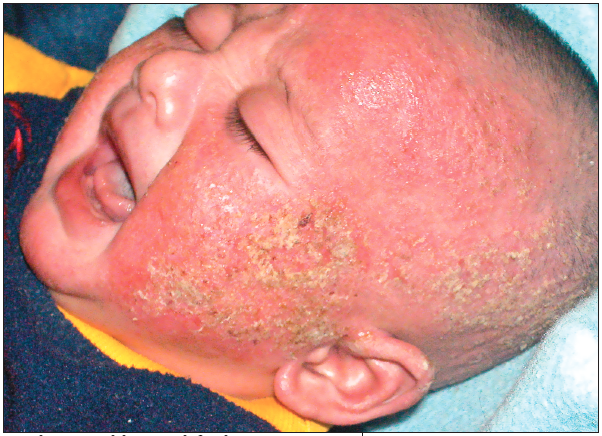 The rash on this 4-week-old girl had appeared 5 days earlier on her face as thickened scales on an erythematous base and subsequently spread to the scalp, shoulder, chest, abdomen, and extremities. A few bullae were noted on the neck and hands. Initial treatment with cephalexin failed to control the rash, and the infant was admitted to the hospital for further evaluation.
The white blood cell count was 14,400/μL (normal range, 5000 to 19,500/μL), with 11% eosinophils. Culture taken from a ruptured bulla grew Staphylococcus sciuri and Enterobacter asburiae. Several antibiotics were tried; S sciuri was resistant to oxacillin and penicillin G, and E asburiae was resistant to amoxicillin and ampicillin. Both organisms were sensitive to gentamicin, ciprofloxacin, and levofloxacin. The infant was treated with gentamicin and was switched to soy-based formula after IgG antibody test results for cow's milk allergen were found to be positive. By day 3, the infant had improved remarkably and was discharged.
The skin of patients with atopic dermatitis may become infected with opportunistic bacteria that prolong healing and may cause serious scarring and morbidities. These infections have been managed empirically, using therapies active against the most common causative organisms—S aureus and methicillin-resistant S aureus.1 However, clinicians who focus only on such common culprits may miss true diagnoses.
S sciuri is an important antibiotic-resistant organism to consider in this setting. It is associated with wound infection, peritonitis, urinary tract infection, pelvic inflammatory disease, splenic abscess, endophthalmitis, endocarditis, and septic shock.2-4 Resistance to macrolides, lincosamides, streptogramins, linezolid, and aminoglycosides has been reported.3,4 E asburiae is a gramnegative rod that has been isolated from various human sources, such as urine, the respiratory tract, wounds, and blood. It has been linked to cases of communityacquired pneumonia.5