Cutaneous Manifestations of HIV Infection in Children Part 1: Infection
The skin is a common site for clinical manifestations of HIV infection. Cutaneous findings may occur at the onset of the infection—or they may indicate progression of the disease with impairment of the patient’s immunity.
Typically, skin disease in HIV-infected children is more severe and more recalcitrant to treatment than in age-matched uninfected controls. Most skin manifestations in infected children fall into 1 of 6 groups:
•Infections.
•Proliferative disorders.
•Vascular disorders.
•Drug eruptions.
•Neoplasms.
•Nutritional deficiencies.
Skin infections and proliferative conditions, such as atopic dermatitis, are most commonly described in children with HIV. They also occur in otherwise healthy patients, of course, but they manifest with atypical severity and distribution and/or at an unusual age in those who have HIV infection. Thus, unusual manifestations of common pediatric skin disorders may be a marker for the presence of HIV.
The following pictures were tak-en of children with various types of fungal, bacterial, and viral infections. None had access to antiretroviral ther-apy. Clearly, the use of antiretro-viral drugs will delay the manifestations and progression of HIV disease.
Part 2 of this Photo Essay, which will appear in the next issue of this journal, will focus on noninfectious com-plications of pediatric HIV infection.

Fungal Infection
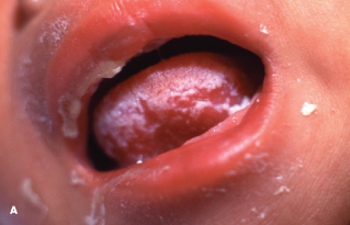
Mucocutaneous candidiasiswas noted in the initial descriptions of pediatric HIV infection in 1983. Most HIV-positive babies have several episodes of oral or perianal candidiasis. Oral candidiasis is the most common fungal infection seen in HIV-infected children. In 20% of patients, this infection progresses to include the esophagus; the result is dysphagia and anorexia. With decreasing immunity, infection becomes more widespread and more resistant to therapy. Infection also tends to recur once treatment is discontinued. Figure A shows a 3-week-old baby with HIV infection and severe oral candidiasis.

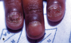
Onychomycosis typically occurs with advanced immunosuppression and may require prolonged courses of oral fluconazole or griseofulvin, depending on the organism involved. Figure B shows a 7-year-old HIV-infected child with long-standing Tricho-phyton rubrum infection of the fingernails.

Viral Infection
In children infected with HIV, herpes simplexlesions may cover large areas of the face; lesions may be multiple or may cause deep ulcerations complicated by bacterial superinfection, scarring, and dyspigmentation. In addition to the oral and nasal mucosa, the eye is a common site of infection. Oral or intravenous acyclovir is usually required to control the disease.
Figure Ashows an 8-year-old HIV-infected girl with severe facial herpes simplex lesions. The lesions were super-infected with Candida albicans and Staphylococcus aureus. Treatment included oral and topical acyclovir, as well as topical antibacterial and antifungal medication. Figure Bshows the residual depigmentation after several weeks of therapy.

Varicella lesions may be hemorrhagic or accompanied by inflammatory hyperkeratosis. The occurrence of herpes zoster in young children raises the specter of underlying HIV infection, intrauterine varicella, or varicella infection at an early age. Lesions can occur in infancy. Figure C shows a 4-year-old child with herpes zoster that involves the left eye.

Herpes zoster in HIV-infected patients can lead to local extensive lesions that become bullous and confluent (Figure D). Chronic herpes zoster is typical in HIV-infected persons and may persist for several months. In severe cases, intravenous acyclovir is indicated. Ophthalmologic evaluation and topical antiviral ophthalmic agents are necessary if the eye is involved or threatened.

Molluscum contagiosumoccurs commonly in HIV disease and may manifest with multiple or giant lesions that are difficult to treat. Figure E shows a 4-year-old HIV-infected child with facial molluscum contagiosum.
Common warts are also frequent and persistent. Figure F shows extensive buccal warts in a 10-year-old boy with advanced HIV disease.

Genital condylomata acuminata can grow to impressive sizes, as in the 4-year-old girl shown in Figure Gand in a 2-year-old boy in Figure H. The lesions in the child in Figure G were surgically removed; the child in Figure H responded to topical therapy.
Bacterial Infection
The most common bacterial skin infections complicating HIV in children are caused by Staphylococcus aureus (impetigo, particularly bullous impetigo; folliculitis; and furuncles), group A b-hemolytic Streptococcus pyogenes (impetigoand lymphadenitis), and Pseudomonas aeruginosa (ecthyma gangrenosumand folliculitis). In all cases, appropriate antibiotic therapy must be instituted without delay.

Figure A shows the extent to which impetigo lesions can occur in children with HIV. Those affected need prompt and vigorous parenteral anti-biotic therapy. Figure B shows the typical lesions of ecthyma gangrenosum. P aeruginosa was cultured from the lesions and the blood of this infant who is HIV-positive.
Mycobacterium tuberculosis skin infection is rare in children. The incidence of atypical mycobacterial infection (Mycobacterium avium complex) in HIV-infected children with a CD4 cell count of less than 100/µL is about 10%. Bacillary angio-matosis caused by Bartonella henselae occurs mostly in adults with advanced HIV disease.


