
An Atlas of Lumps and Bumps, Part 43: Blue Nevi
Blue Nevi
Blue nevi are benign neoplasms composed of dermal melanocytes presenting as papules, nodules or, rarely, plaques of blue, blue-gray, or blue-black color.1 Histologically, the lesion is characterized by a proliferation of spindled and dendritic melanocytes in a focal area of the reticular dermis.
In the majority of cases, blue nevi are acquired and frequently arise in adolescence.1 They can, however, occur at any age or as a congenital lesion.2,3 In addition to the usual sporadic occurrence, congenital blue nevi may be familial or associated with Carney complex also known as NAME (nevi, atrial myxoma, myxoid neurofibromas, ephelides) syndrome or LAMB (lentigines, atrial myxomas, blue nevi) syndrome.4 Blue nevus occurs in approximately 3% of Japanese and 0.5 to 4% of White adults.5 The male-to-female ratio is approximately 1:2.1,4,6
It is believed that a blue nevus results from a dermal arrest in the migration of neural crest melanocytes during fetal development with subsequent proliferation of these melanocytes.7 This is supported by the fact that melanocytes in the blue nevus typically stain positive for Human Melanoma Black-45 (HMB-45).8 The majority of blue nevi have a somatic mutation in the GNAQ or GNA11genes that encode the G-protein α-subunits.9,10 Blue nevi with mutations in KRAS or CYSLTR2 genes have also been reported.9-11 Predisposing factors or triggers include sunburns, trauma, vesiculobullous dermatoses, and oral contraceptives.12,13
Several variants of blue nevi have been described; the two main ones being common blue nevus (also known as dendritic blue nevus) and cellular blue nevus.10 The common blue nevus typically presents as a solitary, smooth, well-demarcated, dome-shaped papule or nodule that measures less than 10 mm in diameter.1,10 The lesion is homogeneously blue to blue-gray to dark-blue in color and is asymptomatic.1,10 The characteristic blue color (ceruloderma) can be attributed to the deep dermal location of melanin which results in the shorter wavelength blue light reflected back to the observer (Tyndall effect).1,10 Sites of predilection include the face (Figure 1), and dorsal surfaces of the hands and feet (Figure 2).10 Rarely, they may occur in the subungual area, conjunctiva, oral mucosa, sino-nasal mucosa, orbit, esophagus, bronchus, lymph nodes, endometrium, uterine cervix, vagina, penis, and prostate.6,7,14,15
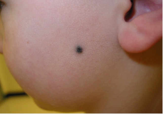
Fig. 1 One of the sites of predilection for blue nevi include the face as shown here.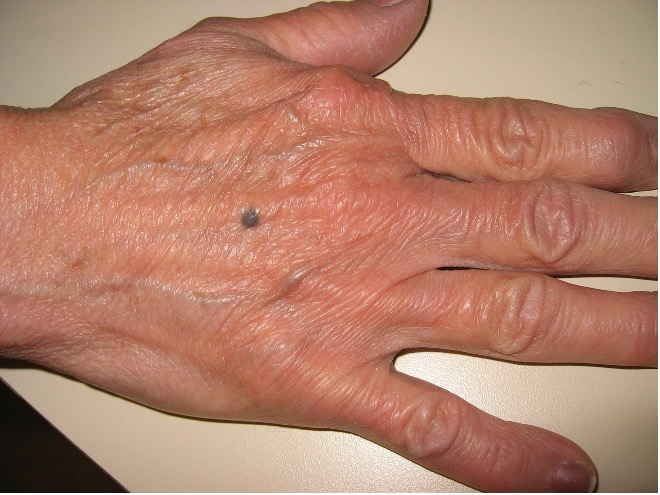
Fig. 2 The lesion is homogeneously blue to blue-gray to dark-blue in color and is asymptomatic.
The cellular blue nevus is less common and is larger (typically 10 to 30 mm in diameter), more elevated, often more heavily pigmented, and has a more irregular surface (Figure 3).10 Sites of predilection include the scalp, face, buttocks, sacrococcygeal region, and extremities.3,4
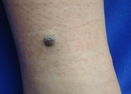
Fig. 3 The cellular blue nevus is a variant of blue nevi and is typically 10 to 30 mm in diameter, more elevated, often more heavily pigmented than blue nevi.
Rarely, blue nevi with peripheral satellitosis have also been described.16 A blue nevus with satellite lesions is an ominous indicator of potential malignant change and should be differentiated from a malignant melanoma with cutaneous metastases.17-19
The plaque-type variant is usually present at birth or in early childhood and may enlarge at puberty.6,20 The lesion is usually blue-gray and composed of a single plaque or a confluence of several small macules, papules and/or nodules.6,21 Sites of predilection include the trunk followed by the scalp.19,21 An important characteristic of a plaque-type blue nevus is the formation of subcutaneous cellular nodules which have the potential to develop into melanoma.20,22
Epithelioid blue nevus is a distinctive variant of blue nevus characterized by a predominately dermal proliferation composed of heavily pigmented epithelioid melanocytes with fewer nonpigmented to minimally pigmented spindled melanocytes.23 Clinically, an epithelioid blue nevus presents as a solitary, heavily pigmented, blue-black, dome-shaped nodule, or as multiple nodules.8 Multiple epithelioid blue nevi have a strong association with Carney complex.8,23
Other rare variants include hypopigmented/amelanotic blue nevus, compound blue nevus, targetoid blue nevus, agminated blue nevus, linear blue nevus, sclerosing blue nevus, eruptive blue nevi, and disseminated blue nevi.3,4,10,13,24-29
The diagnosis is a clinical one based on the characteristic features which can be aided by dermoscopy. Dermoscopy typically shows a bluish or steel-blue, well-circumscribed, homogeneous, structureless pigmentation secondary to the presence of heavily pigmented melanocytes in the dermis.30-32 Polarized dermoscopy may show striking color changes of yellow, pink, green, and purple resembling the colors of a rainbow.33
A blue nevus can be cosmetically unsightly and an esthetic concern if it occurs in an exposed area. A halo phenomenon associated with a blue nevus is rare and the nevus tends to persist unlike the halo phenomenon in other melanocytic nevi.34 Malignant transformation is also rare but should be considered if there is a sudden increase in size of the lesion, deepening of color, asymmetry of the lesion, irregular border, or recurrence after the excision of the lesion.4,35
AUTHORS:
Alexander K.C. Leung, MD1,2, Benjamin Barankin, MD3, Joseph M. Lam, MD4, Kin Fon Leong, MD5
AFFILIATIONS:
1Clinical Professor of Pediatrics, the University of Calgary, Calgary, Alberta, Canada
2Pediatric Consultant, the Alberta Children’s Hospital, Calgary, Alberta, Canada
3Dermatologist, Medical Director and Founder, the Toronto Dermatology Centre, Toronto, Ontario, Canada
4Associate Clinical Professor of Pediatrics, Dermatology and Skin Sciences, the University of British Columbia, Vancouver, British Columbia, Canada.
5Pediatric Dermatologist, the Pediatric Institute, Kuala Lumpur General Hospital, Kuala Lumpur, Malaysia
CITATION:
Leung AKC, Barankin B, Lam JM, Leong KF. An atlas of lumps and bumps, part 43: blue nevi. Consultant. 2024;64(9):eXX.DOI:10.25270/con.2024.10.000004
CORRESPONDENCE:
Alexander K. C. Leung, MD, #200, 233 16th Ave NW, Calgary, AB T2M 0H5, Canada (aleung@ucalgary.ca)
EDITOR’S NOTE:
This article is part of a series describing and differentiating dermatologic lumps and bumps. To access previously published articles in the series, visit: https://www.consultant360.com/resource-center/atlas-lumps-and-bumps.
- Leung AKC, Barankin B. An adolescent with a smooth, blue-black nodule on the dorsal wrist: Blue nevus. Consultant for Pediatricians. 2014;13:501-503.
- Rocha CR, Grazziotin TC, Rey MC, Luzzatto L, Bonamigo RR. Congenital agminated melanocytic nevus--case report. An Bras Dermatol. 2013;88(6 Suppl 1):170-2. doi:10.1590/abd1806-4841.20132137.
- Zembowicz A. Blue nevi and related tumors. Clin Lab Med. 2017;37(3):401-415. doi:10.1016/j.cll.2017.05.001.
- Austad SS, Athalye L. Blue Nevus. In: StatPearls [Internet]. Treasure Island (FL): StatPearls Publishing; 2020 Nov 23-2021 Jan.
- Yoshida Y, Kubota Y, Kiryu H, Nakayama J. Dermoscopic features of blue nevus on the nose. 2006;45(11):1351-2. doi:10.1111/j.1365-4632.2006.02831.x.
- Murali R, McCarthy SW, Scolyer RA. Blue nevi and related lesions: a review highlighting atypical and newly described variants, distinguishing features and diagnostic pitfalls. Adv Anat Pathol. 2009;16(6):365-382. doi:10.1097/PAP.0b013e3181bb6b53.
- Webster TG, McGuinn KP, Del Rosario E, Lee JB, Trufant JW. Subungual blue nevus. J Cutan Pathol. 2020;47(12):1111-1114. doi:10.1111/cup.13781.
- Jang MS, Kang DY, Park JB, Kim ST, Suh KS. Epithelioid blue nevus not associated with Carney complex in a Korean patient. Indian J Dermatol Venereol Leprol. 2012;78(4):521. doi:10.4103/0378-6323.98105.
- Rodríguez-Jiménez P, Mayor-Sanabria F, Rütten A, Fraga J, Llamas-Velasco M. Agminated blue nevus: GNAQ mutations and beyond. Actas Dermosifiliogr. 2021;112(1):95-97. doi:10.1016/j.ad.2019.05.011.
- Shaffer JV, Bolognia JL. Acquired melanocytic nevi (moles). In: Post TW, ed. UpToDate. Waltham, MA. (Accessed on February 28, 2021)
- Möller I, Murali R, Müller H, Wiesner T, Jackett LA, Scholz SL, et al. Activating cysteinyl leukotriene receptor 2 (CYSLTR2) mutations in blue nevi. Mod Pathol. 2017;30(3):350-356. doi:10.1038/modpathol.2016.201.
- Kim YJ, Cohen PR. Blue nevus developing at a combined tetanus, diphtheria, and pertussis (Tdap) vaccination site: Case report and summary of vaccine-associated cutaneous lesions.
- Cureus. 2019;11(6):e4997. doi:10.7759/cureus.4997.
- Maredia H, Eseonu A, Rozati S. Disseminated eruptive blue nevi in a young adult patient. Cureus. 2020;12(11):e11298. doi: 10.7759/cureus.11298.
- Dehavay F, Goettmann S, Zaraa I, et al. Nail unit blue nevi: A 11-case series and review of the literature. Skin Appendage Disord. 2020;6(5):287-295. doi:10.1159/000509204.
- Satolli F, Gandolfi M, Rovesti M, et al. Blue nevus of the nail: A case report and review. Dermatol Ther. 2020 Jul;33(4):e13763. doi:10.1111/dth.13763.
- Yonei N, Kimura A, Furukawa F. Common blue nevus with satellite lesions needs a differential diagnosis from malignant melanoma. Case Rep Dermatol. 2013;5(3):244-247. doi:10.1159/000355179.
- Lourari S, Lamant L, Viraben R, Paul C, Meyer N. Photoletter to the editor: Blue nevus with satellitosis mimicking melanoma. Contribution of dermoscopy and reflectance confocal microscopy. J Dermatol Case Rep. 2012;6(2):54-56. doi:10.3315/jdcr.2012.1097.
- Oliveira AHK, Shiraishi AFMC, Kadunc BV, Sotero PC, Stelini RF, Mendes C. Blue nevus with satellitosis: case report and literature review. An Bras Dermatol. 2017;92(5 Suppl 1):30-33. doi:10.1590/abd1806-4841.20175267.
- Sardoy A, Bidabehere MB, Gubiani ML, Pinardi BA. Blue nevus with satellite lesions mimicking malignant melanoma. Actas Dermosifiliogr. 2018;109(4):377-378. doi:10.1016/j.ad.2017.04.029.
- Furuya A, Namiki T, Takayama K, et al. Dermal melanoma arising in a congenital large plaque-type blue nevus. J Dtsch Dermatol Ges. 2017;15(8):842-844. doi:10.1111/ddg.13275.
- Agusti-Mejias A, Messeguer F, García R, Sánchez JL, Alegre V. Plaque-type blue nevus on the palm. Int J Dermatol. 2013;52(11):1405-1407. doi:10.1111/j.1365-4632.2011.05153.x.
- Yeh I, Fang Y, Busam KJ. Melanoma arising in a large plaque-type blue nevus with subcutaneous cellular nodules. Am J Surg Pathol. 2012;36(8):1258-1263. doi:10.1097/PAS.0b013e31825b62ec.
- Blebea C, Li D, Castelo-Soccio L, Chu EY. Generalized congenital epithelioid blue nevi (pigmented epithelioid melanocytomas) in an infant: Report of case and review of the literature. J Cutan Pathol. 2019;46(12):954-959. doi:10.1111/cup.13544.
- Aydingoz IE, Dikicioglu-Cetin E. Sclerosing cellular blue nevus simulating melanoma. Dermatol Online J. 2012;18(11):11.
- Eichenfield DZ, Cotter D, Thorson J, Hinds B, Sun BK. Agminated blue nevus with a GNAQ mutation: A case report and review of the literature. J Cutan Pathol. 2019;46(2):130-133. doi:10.1111/cup.13373.
- Jaquemus J, Perron E, Buisson A, Ferrara G, Haddad V, de la Fouchardiere A. Compound blue nevus: a reappraisal of the concept in the genomic era. Virchows Arch. 2020;476(3):439-443. doi:10.1007/s00428-019-02667-w.
- Kesty K, Zargari O. Eruptive blue nevi. Indian J Dermatol Venereol Leprol. 2015;81(2):198-201. doi:10.4103/0378-6323.152302.
- Lisboa AP, Silvestre KJ, Pedreira RL, Alves NR, Obadia DL, Azulay-Abulafia L. Agminated blue nevus - Case report. An Bras Dermatol. 2016;91(5):658-660. doi:10.1590/abd1806-4841.20164448.
- Ma C, Chambers CJ, Kiuru M, Marsee DK, Silverstein M. Amelanotic blue nevus. JAAD Case Rep. 2017;3(2):93-94. doi:10.1016/j.jdcr.2017.01.003.
- Di Cesare A, Sera F, Gulia A, et al. The spectrum of dermatoscopic patterns in blue nevi. J Am Acad Dermatol. 2012;67(2):199-205. doi:10.1016/j.jaad.2011.08.018.
- Fei WM, Li CX, Cui Y. Diagnostic value of dermoscopy combined with reflectance confocal microscopy for clinically equivocal blue nevus. Chin Med J (Engl). 2020;133(17):2116-2118. doi:10.1097/CM9.0000000000001007.
- Kim JK, Nelson KC. Dermoscopic features of common nevi: a review. G Ital Dermatol Venereol. 2012;147(7):141-148. PMID: 22481577.
- Uzunçakmak TK, Ozkanli S, Karadağ AS. Dermoscopic rainbow pattern in blue nevus. Dermatol Pract Concept. 2017;7(3):60-62. doi:10.5826/dpc.0703a13.
- Arzberger E, Niederkorn A, Cerroni L, Hofmann-Wellenhof R. Blue nevus with halo? J Am Acad Dermatol. 2016;75(1):e15-16. doi:10.1016/j.jaad.2015.12.057.
- Toledo-Pastrana T, Rodríguez Pérez I, Eguino Gorrochategui P. Malignant blue nevus: A challenge for dermatologists and dermatopathologists. Actas Dermosifiliogr. 2018;109(2):181-183. doi:10.1016/j.ad.2017.02.031.