
An Atlas of Lumps and Bumps, Part 44: Dermatosis Papulosa Nigra
Dermatosis Papulosa Nigra
Dermatosis papulosa nigra is a common, benign epidermal growth characterized by the presence of multiple, asymptomatic, hyperpigmented, round, dome-shaped or flat, papules or macules localized predominately on the face, neck, and upper trunk.1-4 This condition occurs predominantly in dark-skinned individuals (Fitzpatrick skin phototypes IV to VI), most commonly affecting people of Asian and African descent.1,3-5 The incidence in Black adults is between 10% and 30%.3,6 Lesions often begin in adolescence; thereafter the incidence increases with age peaking in the sixth decade.4,7,8 The female-to-male ratio is approximately 2:1.4,8-10
The exact etiology is not known. It is believed that dermatosis papulosa nigra is caused by a defect in the nevoid development of the pilosebaceous follicle.8 There is a genetic predisposition as there is a positive family history in ≥ 50% of affected individuals.2,5,8 As the condition occurs mainly in sun-exposed areas, cumulative ultraviolet exposure play a msajor role in the development of lesions.1,4 Activating mutations in FGFR3 (fibroblast growth factor 3) and PIK3CA (encoding for the catalytic p110 subunit of class 1 phosphatidylinositol-4,5-bisphosphate 3-kinase) genes are involved in the pathogenesis of dermatosis papulosa nigra.5,9,11 Some authors consider dermatosis papulosa nigra to be a variant of seborrheic keratosis in people with dark skin.4,11
Clinically, dermatosis papulosa nigra presents as multiple, asymptomatic, superficial, black or dark-brown, round, macules or more often papules (Figure 1).1,3
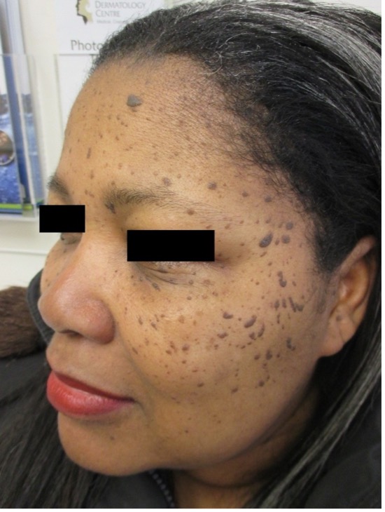
Fig. 1. Dermatosis papulosa nigra presents as multiple, asymptomatic, superficial, black or dark-brown, round, macules or papules.
Lesions are often symmetrically distributed.12 In the early stage, the lesions are often minute and smooth-surfaced. Later, they increase in size and number and become roughened and at times verrucous. Some of the lesions may be filiform or pedunculated. The size of individual lesion usually ranges from 1 mm to 5 mm in diameter and 1 mm to 3 mm in elevation.1,13 Sites of predilection include the face (predominantly the malar regions), neck, upper trunk, and back in descending order of frequency (Figure 2).1,3-5,14 They do not tend to group, and rarely, occur in a linear fashion.15 The lesions do not spontaneously resolve.1,4,16
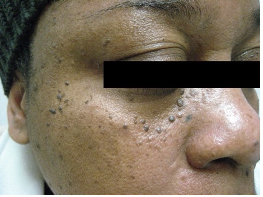
Fig. 2. Sites of predilection include the face, neck, upper trunk, and back.
The diagnosis is mainly clinical. Dermoscopic features include ridges and fissures in a cerebriform pattern, comedo-like openings, and milia-like cysts (Figure 3).4,17 A biopsy should be considered if the diagnosis is in doubt.
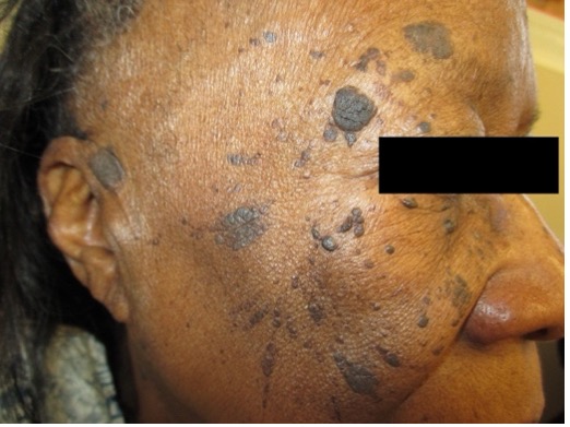
Fig. 3. Dermoscopic features include ridges and fissures in a cerebriform pattern, comedo-like openings, and milia-like cysts.
Dermatosis papulosa nigra can be cosmetically disfiguring and may affect interpersonal relationships.18 In one study, the quality of life of individuals with dermatosis papulosa nigra was moderately affected.6 Other complications include mechanical irritation, and less commonly, inflammation, bleeding, pruritus, and pain.1,13 The condition is generally benign, not related to any systemic disease or syndrome, and without any malignant potential.1,19 However, an abrupt increase in dermatosis papulosa nigra may be a sign of internal malignancy, notably adenocarcinoma of the colon.20
AUTHORS:
Alexander K.C. Leung, MD1,2, Benjamin Barankin, MD3, Joseph M. Lam, MD4, Kin Fon Leong, MD5
AFFILIATIONS:
1Clinical Professor of Pediatrics, the University of Calgary, Calgary, Alberta, Canada
2Pediatric Consultant, the Alberta Children’s Hospital, Calgary, Alberta, Canada
3Dermatologist, Medical Director and Founder, the Toronto Dermatology Centre, Toronto, Ontario, Canada
4Associate Clinical Professor of Pediatrics, Dermatology and Skin Sciences, the University of British Columbia, Vancouver, British Columbia, Canada.
5Pediatric Dermatologist, the Pediatric Institute, Kuala Lumpur General Hospital, Kuala Lumpur, Malaysia
CITATION:
Leung AKC, Barankin B, Lam JM, Leong KF. An Atlas of Lumps and Bumps, Part 44: Dermatosis papulosa nigra. Consultant. 2024;64(10):eXX. doi:10.25270/con.2024.10.000005
CORRESPONDENCE:
Alexander K. C. Leung, MD, #200, 233 16th Ave NW, Calgary, AB T2M 0H5, Canada (aleung@ucalgary.ca)
EDITOR’S NOTE:
This article is part of a series describing and differentiating dermatologic lumps and bumps. To access previously published articles in the series, visit: https://www.consultant360.com/resource-center/atlas-lumps-and-bumps.
References
- Leung AK, Barankin B. Dermatosis papulosa nigra. Enliven: Clin Dermatol. 2015;1(5):008.
- Bruscino N, Conti R, Campolmi P, Bonan P, Cannarozzo G, Lazzeri L, et al. Dermatosis papulosa nigra and 10,600-nm CO2 laser, a good choice. J Cosmet Laser Ther. 2014;16(3):114-116. doi: 10.3109/14764172.2013.854640.
- Kundu RV, Patterson S. Dermatologic conditions in skin of color: Part II. Disorders occurring predominantly in skin of color. Am Fam Physician. 2013;87(12):859-865.
- Xiao A, Muse ME, Ettefagh L. Dermatosis papulosa nigra. In: StatPearls [Internet]. Treasure Island (FL): StatPearls Publishing; 2020 Sep 27-2021 Jan.
- Metin SA, Lee BW, Lambert WC, Parish LC. Dermatosis papulosa nigra: a clinically and histopathologically distinct entity. Clin Dermatol. 2017;35(5):491-496. doi: 10.1016/j.clindermatol.2017.06.001.
- Uwakwe LN, Souza B, Subash J, McMichael AJ. Dermatosis papulosa nigra: A quality of life survey study. J Clin Aesthet Dermatol. 2020;13(2):17-19.
- Babapour R, Leach J, Levy H. Dermatosis papulosa nigra in a young child. Pediatr Dermatol. 1993;10(4):356-358. doi: 10.1111/j.1525-1470.1993.tb00398.x.
- Taylor SC, Averyhart AN, Heath CR. Postprocedural wound-healing efficacy following removal of dermatosis papulosa nigra lesions in an African American population: a comparison of a skin protectant ointment and a topical antibiotic. J Am Acad Dermatol. 2011;64(3 Suppl):S30-S35. doi: 10.1016/j.jaad.2010.11.009.
- Alani A, Natarajan S. First case of dermatosis papulosa nigra in a white child. Clin Exp Dermatol. 2017;42(7):803-805. doi: 10.1111/ced.13186.
- Grimes PE, Arora S, Minus HR, Kenney JA Jr. Dermatosis papulosa nigra. Cutis. 1983;32(4):385-386, 392.
- Hafner C, Landthaler M, Mentzel T, Vogt T. FGFR3 and PIK3CA mutations in stucco keratosis and dermatosis papulosa nigra. Br J Dermatol. 2010;162(3):508-512. doi: 10.1111/j.1365-2133.2009.09488.x.
- Goldstein BG, Goldstein AO. Overview of benign lesions of the skin. In: Post TW, ed. UpToDate. Waltham, MA. (Accessed on March 3, 2021).
- Molinar VE, Taylor SC, Pandya AG. What's new in objective assessment and treatment of facial hyperpigmentation. Dermatol Clin. 2014;32(2):123-135. doi: 10.1016/j.det.2013.12.008.
- Furukawa F, Mizawa M, Shimizu T. Treatment of dermatosis papulosa nigra using a carbon dioxide laser. J Cosmet Dermatol. 2020;19(10):2572-2575. doi: 10.1111/jocd.13309.
- Grimalt R, Happle R. Superimposed segmental dermatosis papulosa nigra. Clin Exp Dermatol. 2020;45(4):521-523. doi: 10.1111/ced.14121.
- Karadag AS, Ozkanli Ş, Mansuroglu C, Ozlu E, Zemheri E. Effectiveness of the pulse dye laser treatment in a Caucasian woman with dermatosis papulosa nigra. Indian J Dermatol. 2015;60(3):321. doi: 10.4103/0019-5154.156447.
- Bhat RM, Patrao N, Monteiro R, Sukumar D. A clinical, dermoscopic, and histopathological study of Dermatosis Papulosa Nigra (DPN) - An Indian perspective. Int J Dermatol. 2017;56(9):957-960. doi: 10.1111/ijd.13633.
- Ali FR, Bakkour W, Ferguson JE, Madan V. Carbon dioxide laser ablation of dermatosis papulosa nigra: high satisfaction and few complications in patients with pigmented skin. Lasers Med Sci. 2016;31(3):593-5. doi: 10.1007/s10103-016-1906-y.
- Veraitch O, Rickaby W, Robson A, Higgins E, Mellerio JE. Early-onset dermatosis papulosa nigra. Br J Dermatol. 2016;174(5):1148-50. doi: 10.1111/bjd.14324.
- Schwartzberg JB, Ricotti CA Jr, Nouri K. Eruptive dermatosis papulosa nigra as a possible sign of internal malignancy. Int J Dermatol. 2007;46(2):186-187. doi: 10.1111/j.1365-4632.2007.02767.x.